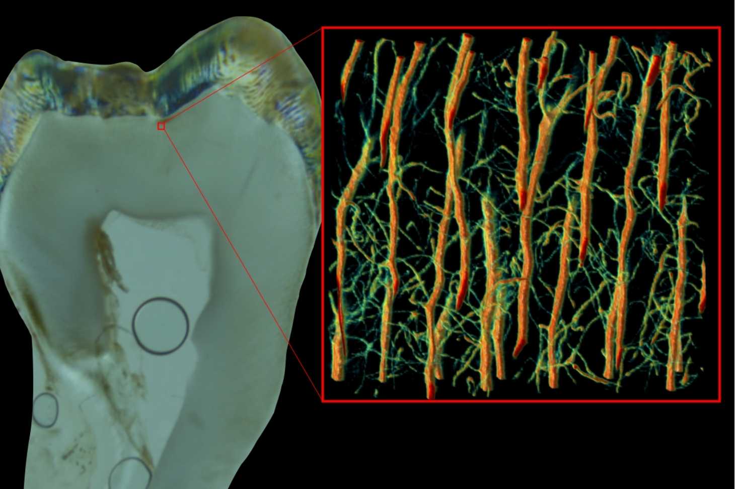
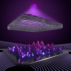
How can the hidden light of living tissue be decoded?
Publication
Research
On November 26, 2025
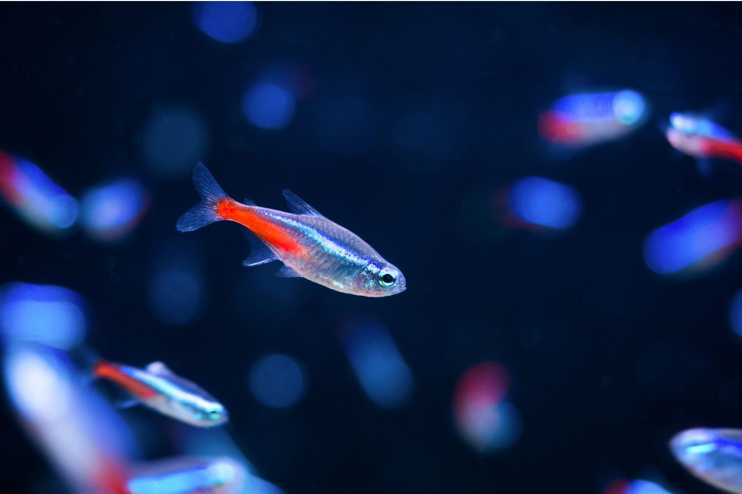
Crédit: KAM Production
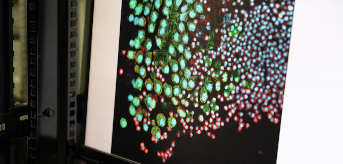
This is the challenge facing the ANR FULBOX project, led by Bastien Arnal, a CNRS researcher at LIPhy, which aims to develop new tools for imaging biological tissues in depth using photoacoustic imaging techniques.
For more information, see:
- the article published in Echosciences Grenoble,
- the video “Concentré de sciences” produced by CNRS Alpes.
Contact
Bastien ARNAL (OPTIMA team)
bastien.arnal univ-grenoble-alpes.fr (bastien[dot]arnal[at]univ-grenoble-alpes[dot]fr)