- Share
- Share on Facebook
- Share on X
- Share on LinkedIn
Séminaire
On September 11, 2023

Magalie Faivre (Institut des Nanotechnologies de Lyon)
It is now well recognized that mechanical signature of cells can give information on their physio-pathological state. Here, we will illustrate this concept by three different approaches able to provide the mechanical phenotype of human cells.
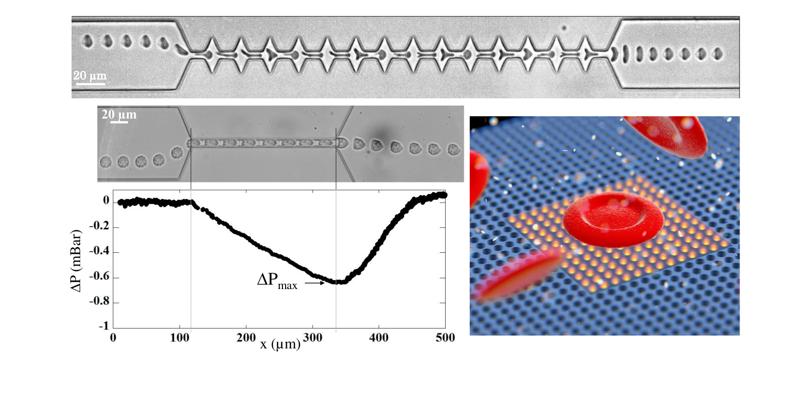
First, we know that the membrane of healthy Red Blood Cells (RBCs) possesses a unique structure responsible of their remarkable ability to deform to go through the small capillaries of the microcirculation. However, various diseases such as malaria [1], Sickle Cell Anemia [2] (SCA) or Hereditary Spherositosis [3] (HS) are associated with impairment of this deformability. We propose to evaluate the possibility to identified mechanically impaired RBCs through their dynamical behaviour when flowing out a microfluidic constriction, hence providing a new type of diagnostic tool based on mechanical phenotyping of cells.
Then, we developed a new bio-photonic approach to perform the characterization of cells deformability. Its originality relies on the all-optical reading of cell deformability using the resonant mode of photonic crystal (PhC) micro-cavities [4]. It has been demonstrated that the presence of an object on top of a PhC micro-cavity induces a local change of refractive index associated with a spectral shift of the resonance spectrum [5]. We propose to extend this concept to the measurement of cell deformability in order to get the mechanical signature of healthy and chemically rigidified RBCs.
Finally, we also addressed the mechanics of more complex cells such as nucleated ones. In Acute myeloid leukemia (AML), a cancer of the myeloid line of blood cells, the abnormal proliferation of leukemia cells build up in the bone marrow and blood, and interfere with normal blood cell production due to their inability to differentiate into mature cells. Recent findings - highlighting an increase of the mechanosensitive pathways in AML relapsed patients [6] - tend to imply a strong interconnection between the cell’s mechanical properties and their ability to survive chemotherapy. We propose to use a passive microfluidic approach [7], quantifying the additional hydrodynamic resistance to the one of the geometric constriction in which they flow, to evaluate the possibility to predict the outcome of a therapeutic treatment using the mechanical signature of AML cell lines. This approach could be the first step towards the development of a new system for personalized medicine.
[1] J.M.A. Mauritz, T. Tiffert, R. Seear, F. Lautenschlger, A. Esposito, V.L. Lew, J. Guck and C.F. Kaminskia, J. Biomed. Opt., 2010, 15(3), 030517.
[2] J.L. Maciaszek and G. Lykotrafitis, J. Biomech., 2011, 44(4), 657-661.
[3] R.E. Waught and P. Agre, J Clim Invest., 1988, 81, 133-141.
[4] L.A. Blanco and M. Nieto-Vesperinas, Journal of Optics A: Pure and Applied Optics, 9(8), (2007), 235–238.
[5] Chow E., Grot A., Mirkarimi L.W., Sigalas M., and Girolami G., Opt. Lett., 29(10), (2004), 1093‑1095.
[6] S. Lefort, unpublished results, 2022.
[7] M. Abkarian, M. Faivre, H.A. Stone, PNAS, 2006, 103, 538-542.
Date
14:00
Localisation
LIPhy, salle de conférence
- Share
- Share on Facebook
- Share on X
- Share on LinkedIn