- Share
- Share on Facebook
- Share on X
- Share on LinkedIn
Recruitment

Nonlinear microscopy is a wide-spread technique for 3D imaging in biological tissues: a pulsed laser is focused into the sample to induce a nonlinear effect such as two-photon excited fluorescence, second or third harmonic generation, in a confined region at the focus, then this focus is scanned to reconstruct a 3D image. Due to the use of near-infrared wavelengths and the intrinsic confinement of the excited volume, nonlinear microscopy is particularly advantageous to look into thick and scattering biological samples.
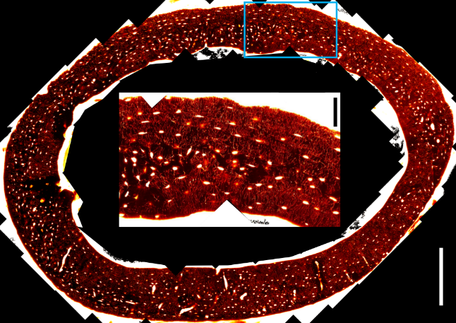
At LIPhy, we are interested in the structure of mineralized tissue, such as bone and teeth, at different scales, from nanometer-sized mineral crystallites to centimeter-sized organs. Optical microscopy has proved a valuable tool to investigate the organization of cellular networks in bone tissues. However, understanding the whole organ structure would require imaging over large field-of-views (cm) and correlating with images obtained by other modalities (such as X-ray or MRI). In most nonlinear microscopes, galvanometric mirrors are used for beam scanning: a deflection of the beam is turned into a translation of the focus by the microscope objective. The characteristics of the optics limits the size of a field-of-view to typically 300 µm and creates spatial distortions. While it is possible to stitch together several images to recover a larger area (see figure), the registration of images from different modalities at the pixel resolution is difficult, due to these distortions.
This project aims at developing a large field-of-view nonlinear microscope with a different scanning pattern. Lateral scanning can be performed over centimeters range by linear translation motors, but motors are relatively slow, compared to galvanometric scanning, so that it would be extremely time-consuming to acquire a stack of images for several axial positions. The idea would be to combine motor-scanning with a fast axial scan at each position of the motors, in order to acquire directly a column of data in the axial direction. This axial scanning is made possible by a tunable acoustic gradient lens (TAG) with a scanning frequency of 70 kHz.
The objectives of this internship would be to test and optimize the optical setup and the acquisition parameters. Then the acquired signal would need to be treated to reconstruct 3D volumes. The microscope will be tested on mineralized tissue samples, in two-photon fluorescence and second-harmonic generation.
We are looking for candidates with a background in optics and physics, and a particular interest in instrumental development and image processing. Knowledge in Python programming would be an asset.
Contact
Irène Wang
OPTIMA team
+33 4 76 51 47 29 – irene.wang univ-grenoble-alpes.fr (irene[dot]wang[at]univ-grenoble-alpes[dot]fr) antoine.delon
 ujf-grenoble.fr ( )
 univ-grenoble-alpes.fr (aurelien[dot]gourrier[at]univ-grenoble-alpes[dot]fr)
- Share
- Share on Facebook
- Share on X
- Share on LinkedIn