- Share
- Share on Facebook
- Share on LinkedIn
Seminar
On June 10, 2024
David Maresca (TU Delft)
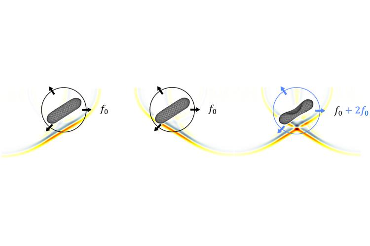
Imaging cellular processes occurring deep within living organisms is fundamental to our understanding of basic biology and disease. In small translucent organisms, fluorescence microscopy can reveal dynamic cellular processes in three dimensions. However, the scattering of light in thick tissue as well as photobleaching of fluorescent proteins limits this method to studying thin specimens. To achieve cellular precision in the context of living opaque organisms, a new imaging paradigm is necessary. I will show that the recent discovery of a “GFP for ultrasound” – genetically encoded gas vesicles that appear bright in ultrasound images – enables to directly connect ultrasound waves to molecular processes of interest. In addition, the combination of gas vesicles with dedicated volumetric ultrasound imaging methods allows for the 3D visualization cellular functions such as gene expression at the cubic centimeter scale.
Contact : Bastien Arnal
Date
14:00
Localisation
LIPhy, salle de conférence
- Share
- Share on Facebook
- Share on LinkedIn