- Imprimer
- Partager
- Partager sur Facebook
- Partager sur LinkedIn
Séminaire
Le 10 juin 2025
Hugues Favre (TU Delft, the Netherlands)
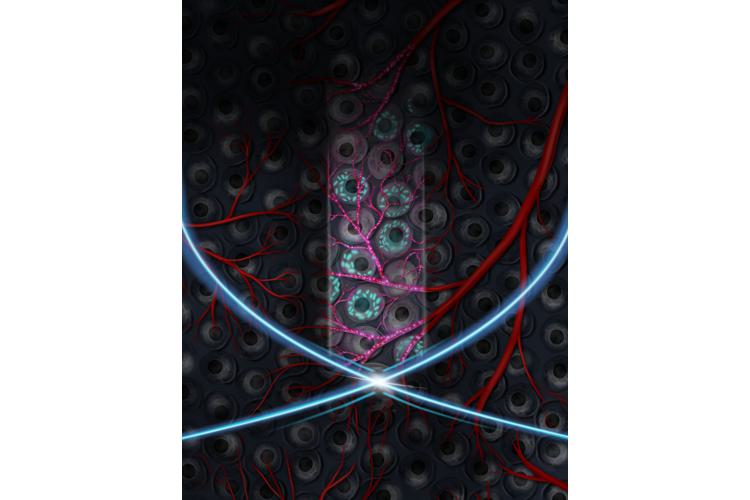
The discovery of the green fluorescence protein (GFP) and the parallel development of super-resolved fluorescence microscopy (2008 and 2014 Nobel Prizes) led to major breakthroughs in basic biology and medicine by enabling the visualization of the pathways of individual molecules inside living cells. While optical imaging is limited to studying thin specimens ( <1mm) due to light scattering in tissue, the introduction of gas vesicles (GVs) as the “GFP for ultrasound” offers an alternative to light for deep-tissue cellular imaging (Bourdeau et al. Nature, 2018). However, current methods bound by the diffraction limit, leading to a resolution of ~100 μm at an ultrasound frequency of 15 MHz. Recently, my laboratory reported the first fast volumetric ultrasound images of acoustic reporter genes based on GVs at the cubic centimeter scale (Heiles et al., Science 2025). While this allows for the detection of cell populations, signals arising from individual cells cannot be isolated. A next frontier in imaging would be to achieve single cell resolution.
In my talk, I will review the fundamentals and theory of nonlinear ultrasound imaging using gas vesicles, which enables live cellular imaging in deep tissue. I will then introduce a new imaging method called nonlinear ultrasound scanning microscopy (nUSM), that can achieve a λ/3 lateral resolution (30 µm). This imaging method represents a significant step toward single-cell resolution, and doubling the transmission frequency could enable the achievement of this goal in future work.
Contact: Bastien Arnal
Date
11:00
Localisation
LIPhy, salle de conférence
- Imprimer
- Partager
- Partager sur Facebook
- Partager sur LinkedIn