- Imprimer
- Partager
- Partager sur Facebook
- Partager sur LinkedIn
Séminaire
Le 21 novembre 2022
Emmanuelle Chaigneau (Institut de la Vision, INSERM)
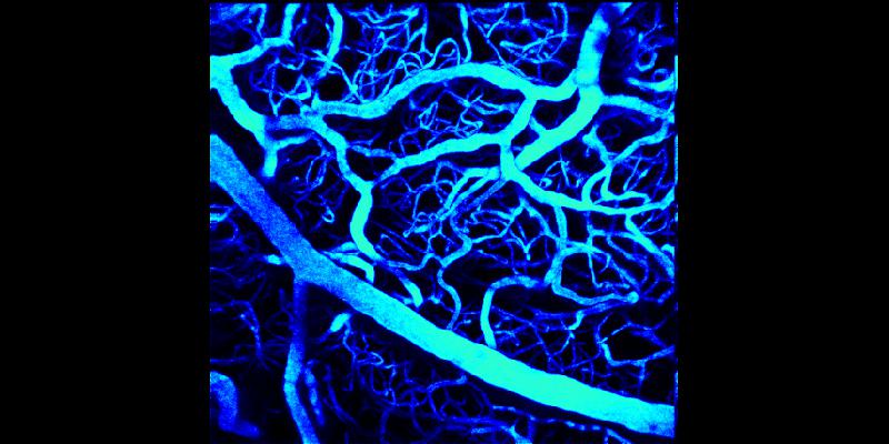
Blood flow is commonly monitored to follow brain activity. At the cellular level, laser scanning microscopy is the technique of choice to measure blood velocity with line-scans and compare sets of experimental conditions. The difference can be as low as a few percent and requires a high precision and reproducibility. Nevertheless, measurements strongly depend on the choice of experimental parameters and image analysis. This is too often ignored causing errors that jeopardize results.
Red Blood Cell (RBC) velocity measurement involves two types of algorithms: The algorithms of the first kind are the image-processing algorithms such as the shear (Kleinfeld et al., 1998), the LSPIV (Kim et al., 2012), the radon-transform (Drew et al., 2010) and the Fourrier transform (Autio et al., 2011;Rungta et al., 2018) algorithms. Each of them provides an apparent velocity (VRBC app) of RBCs in the image. The algorithms of the second kind allow to calculate the real RBC velocity from VRBC app accounting for relativity effects resulting from scanning. They have been ignored for a long time, as it was assumed that the velocity of the scanning mirror largely exceeds the velocity of RBCs. Nevertheless, this assumption only holds true in restricted experimental conditions.
I will first present the algorithm we developed to calculate the real velocity of RBCs and correct for the bias induced by scanning on RBC velocity measurements. I will demonstrate that failing to correct for this relativity effect can result in errors on RBC velocity reaching 100% and show how we validated this algorithm in brain vessels of anesthetized mice (Chaigneau et al., 2019).
Then I will first show how we analyzed the conditions in which some image-processing algorithms can be best used. We developed models that analyze the accuracy of each of the image analysis algorithms. We also developed a software that creates artificial linescan images and used it to validate these models by testing various types of artificial linescan images. Our models predict the experimental conditions in which each image-processing algorithm gives velocity measurements with a given accuracy. I will illustrate the case of different vessel types and give the parameter space available for each of them (Chaigneau et al., 2022).
Overall, our analysis allows unbiased and accurate comparisons of blood hemodynamic parameters from brain capillaries and large vessels in control and pathological animal models.
Date
11:00
Localisation
LIPhy, salle de conférence
- Imprimer
- Partager
- Partager sur Facebook
- Partager sur LinkedIn