- Share
- Share on Facebook
- Share on LinkedIn
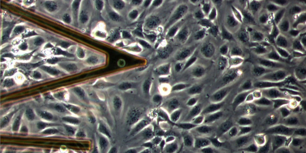
The microscope combines an AFM head and an inverted optical microscope stand allowing to visualize the probe and the studied sample in liquid medium. The system allows to acquire images in transmitted light, phase contrast, or fluorescence. An anti-vibration table minimizes artifacts during sensitive measurements. Finally, it is possible to control the perfusion and the temperature at the sample level thanks to adapted sample holders.
With this device, we can perform the following measurements:
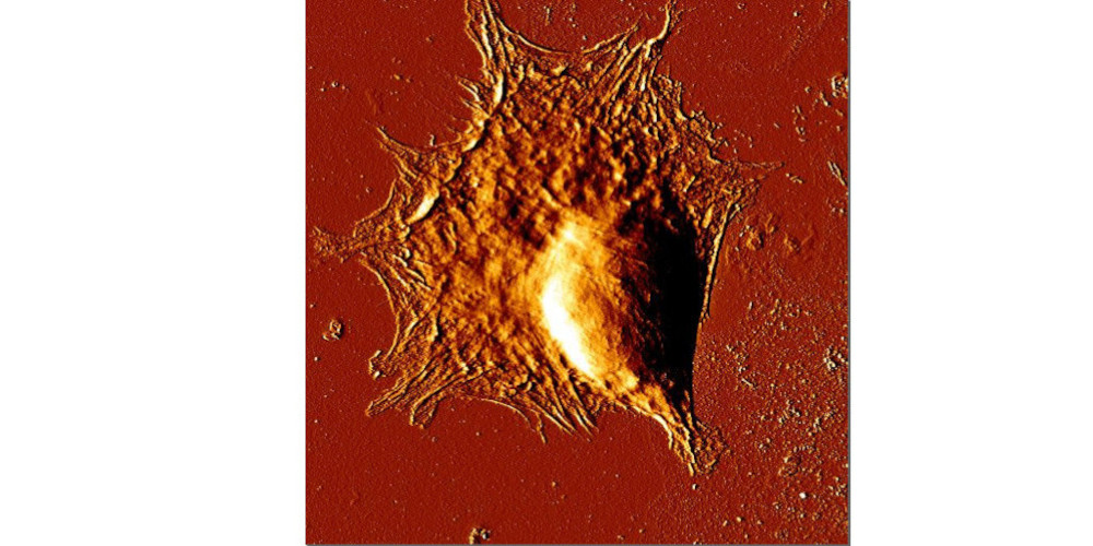
- Topography/relief in scan mode (100µm x 100µm for example)
- Elasticity (force curves, Young's modulus) using Hertz theory with different tip geometries: plane contact, pyramid, sphere
- Viscoelastic properties (1-1000 Hz) in intermittent contact mode (home-made modulus)
- Cell-cell or cell-substrate adhesion, with the complementary CellHesion module (100µm piezo stroke)
Contact
Scientific Leaders
Claude VERDIER
Office 315
04 76 74 84 80
claude.verdier univ-grenoble-alpes.fr (claude[dot]verdier[at]univ-grenoble-alpes[dot]fr)
Valérie LAURENT
Office 313
04 76 74 84 78
valerie.laurentuniv-grenoble-alpes.fr (valerie[dot]laurent[at]univ-grenoble-alpes[dot]fr)
Technical Director
Michaël BETTON
Office 232
04 76 74 83 87
michael.bettonuniv-grenoble-alpes.fr (michael[dot]betton[at]univ-grenoble-alpes[dot]fr)
- Share
- Share on Facebook
- Share on LinkedIn