- Share
- Share on Facebook
- Share on X
- Share on LinkedIn
Thesis defence
On December 13, 2024

Ivana Falco (OPTIMA)
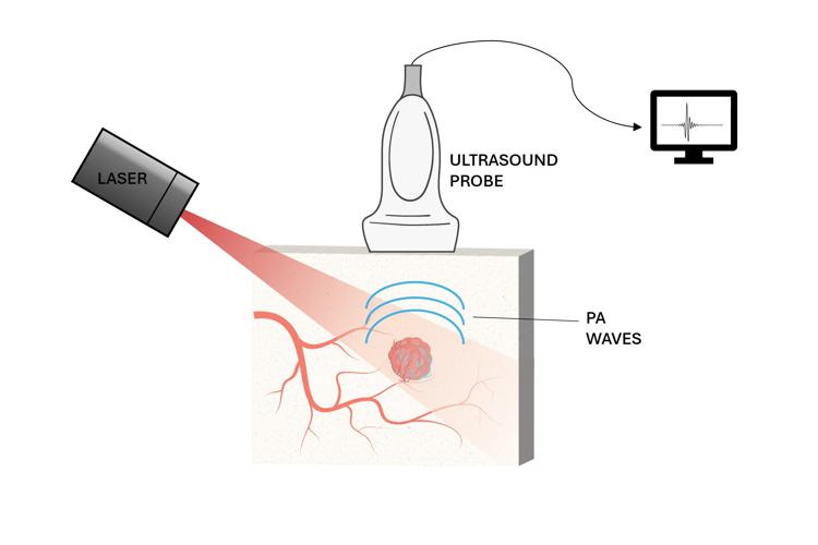
Obtaining images of optical contrast at depths is of significant interest to the biomedical community. A promising approach that offers these advantages, and is the main focus of this manuscript, is Photoacoustic (PA) imaging. Notably, it has the potential to provide real-time imaging at a high spatial resolution and significant penetration depth, targeting endogenous, exogenous, or genetically encoded optical absorbers. Among these, its sensitivity to hemoglobin enables vascular imaging, offering ubiquitous information in biological systems. Moreover, spectroscopic PA acquisitions potentially allow for quantitative imaging of chromophore concentration, which is valuable for a wide range of applications, including blood oxygenation mapping.
For the reasons above, this thesis aims to explore and advance PA imaging by addressing its dual nature – optical and acoustical – each presenting distinct challenges. To this end, we propose innovative approaches where PA imaging is assisted by auxiliary modalities, including both learning-based and imaging-based techniques.
On the acoustical side, the main challenge lies in image reconstruction, which often leads to visibility artifacts that can hide structural details and prevent full analysis. Hence, a part of this study focuses on addressing and correcting these visibility issues. We introduce a method for 3D image reconstruction relying on deep learning. For the first time, by use of a 3D spherical sparse array, measurable artifact-free images, known as Photoacoustic Fluctuation images (PAFI), are used as ground truths to train a convolutional neural network. This approach allows for real-time correction of single-shot PA images and its effectiveness is demonstrated on a dataset of chicken embryo model vasculature and in preliminary in vivo experiments in mice.
The other key focus of this thesis is addressing the optical challenges towards quantitative PA imaging. Accurate light fluence estimation is crucial for quantitative analysis, and it is intrinsically linked to the optical properties of tissues. In the related studies presented in the manuscript, ultrasound power Doppler (UPD) imaging is coupled with PAFI imaging, as both are sensitive to blood vessels and share the same acquisition system. Specifically, we propose to combine the blood information from PAFI and UPD to uniquely and experimentally determine the 3D light fluence distribution within blood vessels. This approach is validated through numerical simulations and experiments using flow phantoms in light-scattering media.
Beyond direct fluence measurement, this study validates that UPD carries information about the effective fractional volume of blood at the imaging resolution, serving as the foundation for a second numerical investigation. In this latter, UPD is employed as a blood prior in a diffuse optical PA inverse problem to mitigate the issue of non-uniqueness of the solution, ultimately converging to accurate optical properties of tissues.
Date
13:30
Localisation
LIPhy, salle de conférence
- Share
- Share on Facebook
- Share on X
- Share on LinkedIn