- Share
- Share on Facebook
- Share on X
- Share on LinkedIn
Thesis defence
On November 25, 2024

Ali Wahhod (MC2)
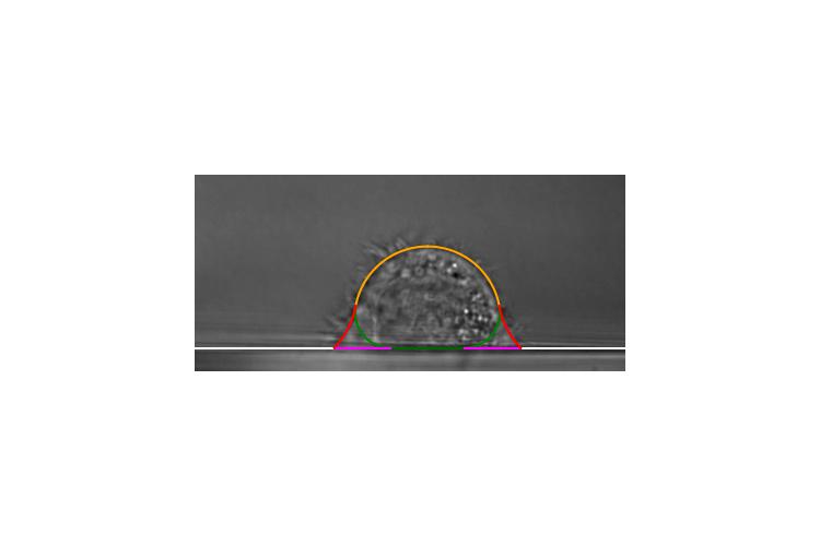
Cell shape is intricately connected to cytoskeleton mechanics, particularly the actomyosin cortex—a gel-like structure located beneath the cell membrane. Myosin-driven mechanical tension in this cortex can be modelled as surface tension, which helps shape the cell as it spreads on a substrate, often resembling a spherical cap. However, the reversal in surface concavity near the triple line with the substrate suggests that the actomyosin cortex alone does not fully govern the cell’s mechanical balance.
Using fluorescence microscopy, we identified a vimentin cortex beneath the actomyosin layer, suggesting that vimentin also plays a key role in regulating cell shape. To explore this further, we modeled the cell’s spreading behaviour by solving the Young-Laplace equation, viewing the interfaces shaped by the actomyosin and vimentin cortices as surfaces of constant mean curvature (nodoids and unduloids).
Through an inverse problem approach, we successfully reconstructed the 3D geometry of the cell based on geometric data such as contact radius, equatorial radius, and cell height. However, while this reconstruction provided valuable insights, the lack of force measurement in the this system limited our ability to calculate absolute tensions—allowing us to only determine relative tension values. This limitation led us to use data obtained with a parallel-plate system, which offers direct force measurements. These measurements enable us to determine absolute tensions and pressures, overcoming the constraints of relative tension calculations. Our results suggest that the vimentin cortex maintains different mechanical pressures in distinct cellular compartments, likely driven by osmotic pressure differences.
Date
14:00
Localisation
LIPhy, salle de conférence
- Share
- Share on Facebook
- Share on X
- Share on LinkedIn