- Share
- Share on Facebook
- Share on LinkedIn
Thesis defence
On December 19, 2025
Lauren Anderson (OPTIMA, LIPhy)
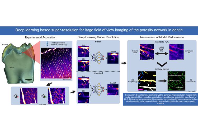
Imaging dentinal porosity is currently a challenging topic in dental research. This porosity results from the presence of cellular processes from odontoblasts, located in the pulp and believed to play a key role in mechano-sensing. It consists of microscopic tubules, interconnected with sub-microscopic branches containing cell processes surrounded by physiological fluids. The recent observation that these pore vessels form a dense network led to the realization that mechanical or thermal stimuli propagation could be more complex than currently thought. Acquiring an accurate 3D representation of this network is therefore key to understanding tooth function. This imposes strong constraints on imaging resolution, which needs to be in the 100 nm range to visualize the smallest pores, while achieving visualization of an entire tooth section to take into account histology and anatomical fluctuations. This is currently beyond the reach of any existing imaging modality or instruments.
To achieve high-resolution (HR) visualization on the full tooth scale, we propose using deep learning (DL) super-resolution (SR) models trained on confocal fluorescence microscopy images, the current gold-standard for 3D visualization of dentinal porosity. The principle is to apply SR models to restore the highest achievable optical resolution for a given microscope on degraded images acquired much faster by under-sampling the scan. This is achieved by training SR models using pairs of low-resolution (LR) and HR images. Three supervised DL models (RCAN, FSRCNN, pix2pix) and one unsupervised model (CycleGAN) were selected and applied to a unique set of experimentally paired high- and low-resolution confocal images acquired with a pixel size increase of x2, x4, and x8. Training was performed on input patches, carefully selected to balance representation of different porosity feature classes. Qualitative assessment on model performance showed good results for all models at x2 degradation and CycleGAN up to x8 degradation. Quantitative analysis of generated HR images was then performed with a set of standard similarity and distribution-based image quality assessment (IQA) metrics, which produced contradictory results compared to visual assessment. This drew the need for the development of a biology-driven evaluation based on scales, morphology, and connectivity of the porosity network. A connected component analysis of 2D scales and morphology followed by a 3D graph network analysis of connectivity was performed. Based on a combination of all quantitative results, CycleGAN and pix2pix models showed good performance up to x8. CycleGAN was selected and improved with new training on an enriched dataset, with the goal of training a more generalizable model. Finally, application of this newly trained model on large FOV LR acquisitions at x4 and x8 was performed, producing 5.8 mm x 3.3 mm x 20.4 µm HR volumes.
Results from this study showed great promise for the use of SR models with confocal microscopy to restore HR information from LR acquisitions. The CycleGAN and pix2pix showed good performance up to x8, which decreased scan time by a factor of 20.3. The biology-driven assessment allowed for a better interpretation of SR model performance specific to dentin porosity features. For example, to identify specific missing or false positive components, the effects of low intensity features, and non-linearities in model generation. With an improved and more generalizable CycleGAN, a large HR FOV could be generated which, including scan-time and model application, provided HR outputs 8.1x faster than a standard HR acquisition and reduced scan time by over 300 hours. Results from this study introduces a great potential for achieving large-scale HR visualization of the microscopic and sub-microscopic porosity network in dentin, working towards imaging dentin porosity in a whole tooth.
Date
15:00
Localisation
LIPhy, conference room
- Share
- Share on Facebook
- Share on LinkedIn