
- Imprimer
- Partager
- Partager sur Facebook
- Partager sur LinkedIn
Séminaire
Le 20 octobre 2026
Fariha Hasan - (PhD, MC2) || Carlos Arauz Moreno - (Post-Doc, IMOV)
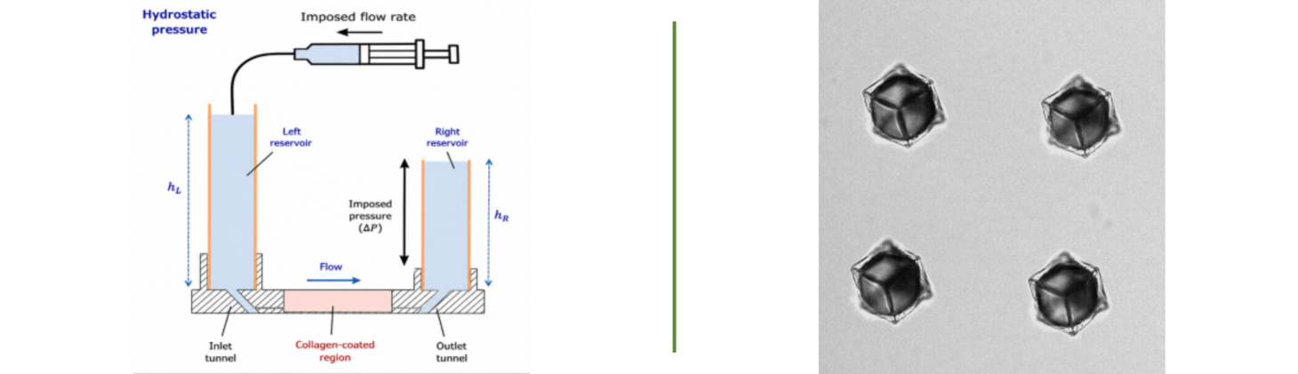
Investigating activity and anisotropy in endothelial monolayers using an optimized microstretcher-on-chip platform.
Fariha Hasan - (PhD, MC2)
The vascular endothelium is continuously exposed to mechanical and geometrical constraints that influences the organization, morphology and collective behavior of cells. To understand the endothelial tissues response to these physical constraints is important to understand the vascular development mechanisms, homeostasis and disease. This project was designed to develop and optimize a biomimetic microvessel-on-chip platform (microstretcher) for studying the effect of geometrical confinement leading to mechanical constraints on the organization of an endothelial monolayer. An optimized and detailed experimental protocol was designed, including the preparation of PDMS microdevices, extraction of collagen from rat tails tendons, preparation of collagen hydrogels, formation of microchannels using needle template, endothelialization (seeding) with Human Umbilical Vein Endothelial Cells (HUVECs), and fluorescent imaging using confocal microscopy. Several optimizations were used to improve the protocols and processes of device fabrication, collagen quality, stability of the hydrogels, endothelial cell viability and endothelial cell monolayer formation. The improvements enabled the reproducible creation of physiologically sized endothelialized microvessels with reproducible diameters. Microvessels of 120 μm and 200 μm diameter were used to see the effect of curvature and geometry on endothelial organization. Cellular morphology and collective organization were quantified with values such as orientation, aspect ratio, circularity, elongation and alignment index, by using confocal microscopy and image processing workflows using ImageJ. The observations indicate that vascular curvature and containment significantly shape the architecture of the endothelial cells and the organization of cytoskeleton.
Resonating caged bubbles at the micrometer scale.
Carlos Arauz Moreno - (Post-Doc, IMOV)
Caged bubbles, i.e., bubbles constrained within a solid (and hollow) cage of arbitrary shape, have been shown to work as affective probes for near-field acoustic imaging in the kHz (mm) range. The reason being their sub-wavelength dimensions (and associated resonance) and the ability of the cage to be manipulated in 3D space. In this talk, I will discuss caged bubbles in the micrometer range, their fabrication (and challenges thereof), and ultimately, their resonant behavior.
Date
10:30
Localisation
LIPhy, salle de conférence
- Imprimer
- Partager
- Partager sur Facebook
- Partager sur LinkedIn