- Share
- Share on Facebook
- Share on LinkedIn
Thesis defence
On November 21, 2025
Seunghwan Lee (OPTIMA, LIPhy)
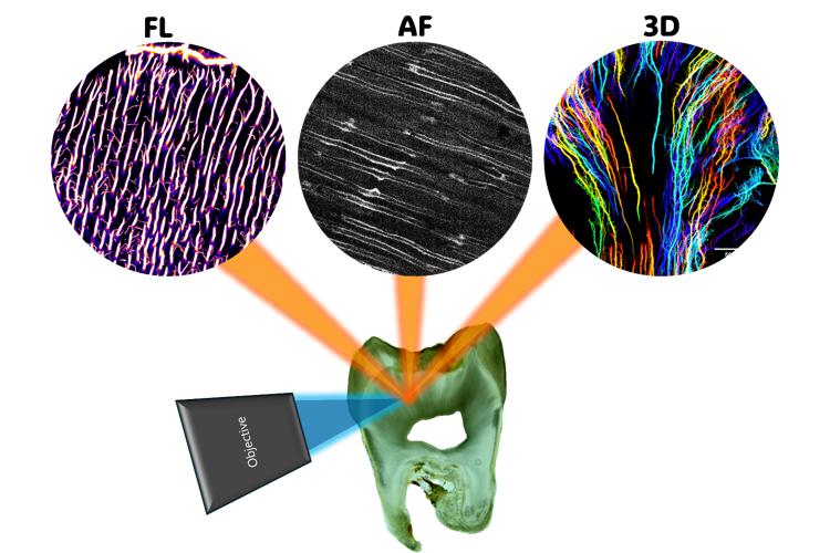
This study aims to establish a methodology to enhance three-dimensional (3D) visualisation of dentin porosity. Dentin is a key feature to understand the mechanical and biological properties of human teeth and could serve as a pathological indicator. Despite its significance, the 3D structure of dentin porosity remains poorly described due to the technical challenges of volumetric imaging with nanometre-scale resolution and the strong light scattering properties of dentin. To overcome these challenges, we developed a protocol that combines sub-wavelength sample surface preparation, refractive index-matched staining, and two-photon microscopy (TPM). The protocol was evaluated by expert visual assessment, using the contrast between porosity and surrounding tissue, as well as optical aberration effects as discriminating criteria. Subsequently, we developed image quality analysis metrics that reflect expert perception. Overall, our study provides a quantitative framework to assess dentin porosity imaging depth limits and an approach that achieves an 8-fold greater imaging depth compared with standard confocal laser scanning microscopy (CLSM). Another focus of our research is demonstrating the value of autofluorescence imaging for dentin porosity. In elderly patients, mineral crystal deposition causes porosity occlusion, which impedes staining medium infiltration. Simultaneous autofluorescence acquisition enabled visualisation of the occluded porosity without additional staining or instrumentation. We further demonstrated the applicability of our 3D porosity analysis through a case study of a rare disease: dentinogenesis imperfecta (DI). A tooth from a patient with type I DI was processed with our sample preparation protocol. Pathological dentin porosity was mapped across the entire dentin using CLSM, and its 3D structure was further visualised in selected regions by TPM. Structural abnormalities of dentin porosity revealed new insights into pathological dentinogenesis in DI. These advances highlight the potential of our protocol for dentin porosity studies in future dental research.
Date
10:00
Localisation
LIPhy, conference room
- Share
- Share on Facebook
- Share on LinkedIn