- Share
- Share on Facebook
- Share on X
- Share on LinkedIn

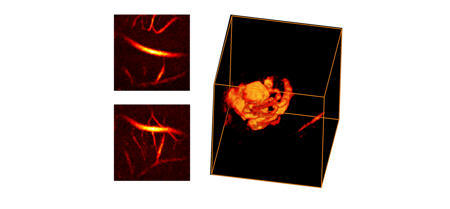
Photoacoustic imaging allows the formation of optical contrast images, deep in the tissue, beyond the limits of optical scattering. When a nanosecond pulse of light is sent into a tissue, the scattered photons will, upon encountering a chromophore, generate a very brief heating, which by thermo-elastic effect, generates an ultrasound wave. After propagation, the measurement of the acoustic waves on the surface of the tissue provides an optical contrast image with acoustic resolution (typically 100 µm). We are developing new systems and methods for image enhancement with the aim of introducing new diagnostic tools in the clinical field.
- Hybrid 3D photoacoustic/ultrasound imager (B. ARNAL, E. BOSSY)
- Super-resolution (B. ARNAL, E. BOSSY)
- Improved imaging of blood vessels (B. ARNAL, E. BOSSY)
- Full field optical vibrometer (O. JACQUIN)
- Photoacoustic arthroscopy of the meniscus (O. JACQUIN, E. LACOT)
- Inverse problems for oxygenation quantification by multispectral photoacoustic imaging (B. ARNAL)
Supervisor
Bastien ARNAL
Emmanuel BOSSY
Olivier JACQUIN
Eric LACOT
Support technician
Sylvie COSTREL
Philippe MOREAU
- Share
- Share on Facebook
- Share on X
- Share on LinkedIn